Past MidWinter Meetings
49th Annual MidWinter Meeting
San Juan, PR - February 7-11, 2026
48th Annual MidWinter Meeting
Orlando, FL - February 22-26, 2025
47th Annual MidWinter Meeting
Anaheim, CA - February 3-7, 2024
46th Annual MidWinter Meeting
Orlando, FL - February 11-15, 2023
45th Annual MidWinter Meeting
Virtual - February 5-9, 2022
44th Annual MidWinter Meeting
Virtual - February 20-24, 2021
43rd San Jose, CA
January 25-29, 2020
42nd Baltimore, MD
February 9-13, 2019
41st San Diego, CA
February 10-14, 2018
40th Baltimore, MD February
11-15, 2017
39th San Diego, CA
February 20-24, 2016
38th Baltimore, MD
February 21-25, 2015
37th San Diego, CA
February 22-26, 2014
36th Baltimore, MD
February 16-20, 2013
35th San Diego, CA
February 25-29, 2012
34th Baltimore, MD
February 19-23, 2011
33rd Anaheim, CA
February 6-10, 2010
32nd Baltimore, MD
February 14-19, 2009
31st Phoenix, AZ
February 16-21, 2008
30th Denver, CO
February 10-15, 2007
29th Baltimore, MD
February 5-9, 2006
28th New Orleans, LA
February 11-15, 2005
27th Daytona Beach, FL
February 21-26, 2004
26th Daytona Beach, FL
February 22-27, 2003
![]()
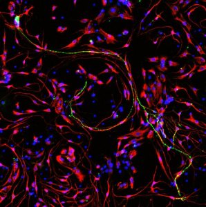 Image courtesy of Patrick Lam, Ella Trang, Niki Gunewardene, and Andrew Wise of Bionics Institute Description: 3-day culture of primary spiral ganglion cells isolated from 5D neonatal Sprague Dawley rats.This image shows a common phenomenon of spiral ganglion neuron processes being surrounded by glial cells that could play an important role in the nerve survival and signalling.Cells were visualised via immunocytochemistry. Labelled in green are two spiral ganglion neurons using Tuj1 as the primary antibody binding marker; in red are the glial cells using S100 beta as the primary antibody binding marker; all cell nuclei were stained with DAPI in blue.