Top Menu

D. Kent Morest, M.D.

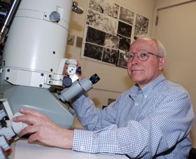
Kent Morest, M.D., (1934-2020)

With profound sadness, we announce the passing of Donald Kent Morest, MD, age 86, on December 30, 2020, in Arlington, MA. Kent was a pioneer in both auditory and developmental neuroscience. He is often regarded as the father of modern auditory neuroanatomy, following in the footsteps of his muse Santiago Ramón y Cajal in exploiting the Golgi method, while also being the first to apply systematic electron microscopic analysis to studies of the auditory system. In addition, his detailed analyses of developing auditory brainstem and other parts of the brain led to important insights on neuronal migration and differentiation.

Kent was born in Kansas City, MO, the son of a physician, which may explain his early interest in medicine. In 1955, Kent received a Bachelor of Arts degree with honors from the University of Chicago. At Chicago, Kent was exposed to basic philosophical questions and an analytical approach that propelled his keen interest in research in the basic sciences. In 1960, he received his MD with honors from Yale University School of Medicine. During and after medical school, he conducted research in several internationally-known laboratories, including the Montreal Neurological Institute, University College London, and the Laboratory of Grant Rasmussen at NIH. After studying with Dr. Rasmussen, Kent decided to pursue basic neuroscience, rather than clinical science. By the age of 31 he was renowned both nationally and internationally for his work describing the organization of the medial geniculate body in the thalamus. During his time in the Rasmussen laboratory, he met Rosemary Richtmyer, who became his wife and partner in the laboratory. Together they developed an unparalleled collection of Golgi-impregnated brain sections, which formed the basis of many important studies, performed with his students and outside collaborators. 

Kent’s first faculty position was at the University of Chicago (1963-1965), followed by an appointment in the Department of Anatomy at Harvard Medical School, where he remained for 12 years, rising through the academic ranks. At Harvard, his lectures to medical and graduate students were outstanding for their intensity, scholarship, and attention to detail. In departmental seminars, he was notorious for asking the critical question. His lab at Harvard bustled with a medley of students at all stages of their careers. There were also joint projects with scientists from the Eaton-Peabody Laboratory at the Massachusetts Eye and Ear Infirmary related to Kent’s long-standing, productive collaboration with its head, Dr. Nelson Kiang. Most remarkable and important to his students were Kent’s weekly lab meetings, where their ongoing research was critiqued. In 1977, Kent accepted a professorship at the University of Connecticut Health Center in Farmington, CT, where he served in the Department of Anatomy (1978-2000) and the Department of Neuroscience (2000-2012). There, he spearheaded many advances in cellular and systems neuroscience, especially auditory neuroscience.

Kent’s interest in the auditory system emerged during his fellowship years in the Rasmussen laboratory. He applied the rapid Golgi method and in subsequent years combined his Golgi studies with electron microscopy and silver degeneration to perform in-depth analyses of the neuronal architecture and synaptic organization of the major components of ascending auditory systems in the brainstem, including the dorsal and ventral cochlear nuclei, medial nucleus of the trapezoid body, inferior colliculus, and medial geniculate body. This work set the stage for physiologists to investigate central auditory signal processing at the cellular level of analysis that was the envy of other sensory system scientists. With his students, he also elucidated the innervation patterns and synaptic organization of the cochlea using both the rapid Golgi method and electron microscopy. Later, Kent applied immunocytochemical methods for key neurotransmitters, including glutamate, GABA, and glycine, to define more fully the synaptic organization and connections of the cochlear nuclei and superior olivary complex. 

At UConn, Kent focused on characterizing the degenerative process in the dorsal cochlear nucleus at the cellular and molecular levels following overstimulation of the cochlea.  Using patterns of axonal degeneration, he documented that peripheral auditory lesions lead to synaptic degeneration in the brain. His studies investigated the balance between excitation and inhibition of synaptic terminals in the cochlear nuclei, focusing on factors that protect or allow synapses to recover normal function after noise-induced damage. His most recent immunocytochemical studies on the auditory system included defining the expression of potassium and sodium channel subunits that change following deafferentation. Thus, throughout his career, Kent contributed cutting-edge findings that combined state-of-the-art approaches with classical neuroanatomical methods to advance our understanding of auditory system structure and function.

Kent’s work on the auditory system alone would have been sufficient for an extraordinary career. Yet, he also produced a remarkable series of papers on neuronal development, focusing on neuronal migration and differentiation of axons, dendrites and synaptic terminals in the cerebral cortex, retina, vestibular and auditory brainstem nuclei, optic tectum, and vestibular and auditory parts of the inner ear. Applying the rapid Golgi method and electron microscopic methods to understand these developing systems, he provided strong support for the concept of perikaryal translocation as a mechanism underlying neuronal migration from the site of proliferation of new neurons to their final destination in the brain. He reconfirmed the importance of growth cones for identifying growing axons and dendrites, and retraction clubs for the withdrawal of improper outgrowths. More recently, he developed innovative cultures of the cochleovestibular ganglion and brainstem to investigate the role of trophic interactions on neuronal development and maintenance. He identified the importance of basic fibroblast growth factor, brain-derived neurotrophic factor, and cell adhesion molecules in this system. In this work, he challenged developmental neurobiologists by demanding that any interpretation of a neurobiological process be based on combined structural and functional data.

One of Kent’s lasting gifts to his students was in sharing his love of the written word. He pored over every word in every manuscript, insisting that the writing be both elegant and evocative. He loved to find just the right word to characterize an anatomical structure, with the goal of bringing life to its description. His efforts to evoke the beauty of anatomical structures extended to his figures. He hoped to help the reader “gain some impression of what the impregnated cells looked like to the microscopist.” Most important, he recognized that it is the “character of the neuropil” that is crucial for understanding the structural basis for integrating inputs within different parts of the nervous system, so the neuropil became a major focus in many of his studies and illustrations.

Kent published 72 full-length papers in auditory neuroanatomy and 38 papers in developmental neurobiology. This is a prolific record, given the high level of detail found in every manuscript. He also coauthored 13 book chapters and two brain atlases. In addition to his dedication to scholarly work, he had a long history of serving with the highest distinction on study sections for NIH, NASA and NSF, editorial boards for prominent journals, and various international committees.  At UConn, he founded the neuroscience area of concentration in the Biomedical Science PhD program, serving as director from 1982 to 1995. He established the Center for Neuroscience that was the predecessor of the current Department of Neuroscience and served on all major committees at UConn. Among other honors, in 1992, he was elected to the Connecticut Academy of Sciences and Engineering, and in 1992-1995, he served on the prestigious Advisory Board of the National Institute for Deafness and other Communication Disorders. According to his family, he was most proud of a Professional Achievement Citation from the University of Chicago, awarded in 2009.

In closing, Dr. D. Kent Morest was a distinguished scientist and recognized leader in the fields of neuroanatomy of the auditory system and developmental neurobiology, with a record of sustained productivity and impeccable scholarship in both. He also was a superb teacher and mentor.  Former students and colleagues deeply admired his encyclopedic knowledge, quick wit, and pervasive demand for excellence. A personal obituary appeared in the Hartford Courant, January 10, 2021.


Written by:

Kenna Peusner, PhD                                       Douglas L. Oliver, PhD

Professor of Neurology                                  Professor and Vice-Chair of Neuroscience

George Washington University                      University of Connecticut School of Medicine

School of Medicine


Sonal Jhaveri, PhD                                         Leslie P. Tolbert, PhD

Science Program Director                              Regents Professor Emerita in Neuroscience

Student Affairs Office                                    University of Arizona

Dana-Farber Cancer Institute


Nell B. Cant, PhD,

Emeritus Faculty

Duke University Medical School

Hearing loss can significantly disrupt the ability of children to become mainstreamed in educational environments that emphasize spoken language as a primary means of communication. Similarly, adults who lose their hearing after communicating using spoken language have numerous challenges understanding speech and integrating into social situations. These challenges are particularly significant in noisy situations, where multiple sound sources often arrive at the ears from various directions. Intervention with hearing aids and/or cochlear implants (CIs) has proven to be highly successful for restoring some aspects of communication, including speech understanding and language acquisition. However, there is also typically a notable gap in outcomes relative to normal-hearing listeners. Importantly, auditory abilities operate in the context of how hearing integrates with other senses. Notably, the visual system is tightly couples to the auditory system. Vision is known to impact auditory perception and neural mechanisms in vision and audition are tightly coupled, thus, in order to understand how we hear and how CIs affect auditory perception we must consider the integrative effects across these senses.

We start with Rebecca Alexander, a compelling public speaker who has been living with Usher’s Syndrome, a genetic disorder found in tens of thousands of people, causing both deafness and blindness in humans. Ms. Alexander will be introduced by Dr. Jeffrey Holt, who studies gene therapy strategies for hearing restoration. The symposium then highlights the work of scientists working across these areas. Here we integrate psychophysics, clinical research, and biological approaches, aiming to gain a coherent understanding of how we might ultimately improve outcomes in patients. Drs. Susana Martinez-Conde and Stephen Macknik are new to the ARO community, and will discuss neurobiology of the visual system as it relates to visual prostheses. Dr. Jennifer Groh’s work will then discuss multi-sensory processing and how it is that vision helps us hear. Having set the stage for thinking about the role of vision in a multisensory auditory world, we will hear from experts in the area of cochlear implants. Dr. René H Gifford will discuss recent work on electric-acoustic integration in children and adults, and Dr. Sharon Cushing will discuss her work as a clinician on 3-D auditory and vestibular effects. Dr. Matthew Winn will talk about cognitive load and listening effort using pupillometry, and we will end with Dr. Rob Shepherd’s discussion of current work and future possibilities involving biological treatments and neural prostheses. Together, these presentations are designed to provide a broad and interdisciplinary view of the impact of sensory restoration in hearing, vision and balance, and the potential for future approaches for improving the lives of patients.

Kirupa Suthakar, PhD - Dr Kirupa Suthakar is a postdoctoral fellow at NIH/NIDCD, having formerly trained as a postdoctoral fellow at Massachusetts Eye and Ear/Harvard Medical School and doctoral student at Garvan Institute of Medical Research/UNSW Australia.  Kirupa's interest in the mind and particular fascination by how we are able to perceive the world around us led her to pursue a research career in auditory neuroscience.  To date, Kirupa's research has broadly focused on neurons within the auditory efferent circuit, which allow the brain to modulate incoming sound signals at the ear.  Kirupa is active member of the spARO community, serving as the Chair Elect for 2021.


I began studying the vestibular system during my dissertation research at the Università di Pavia with Professors Ivo Prigioni and GianCarlo Russo. I had two postdoctoral fellowships, first at the University of Rochester with Professor Christopher Holt and then at the University of Illinois at Chicago with Professors Jonathan Art and Jay Goldberg.

My research focuses on characterizing the biophysics of synaptic transmission between hair cells and primary afferents in the vestibular system. For many years an outstanding question in vestibular physiology was how the transduction current in the type I hair cell was sufficient, in the face of large conductances on at rest, to depolarize it to potentials necessary for conventional synaptic transmission with its unique afferent calyx.

In collaboration with Dr. Art, I overcame the technical challenges of simultaneously recording from type I hair cells and their enveloping calyx afferent to investigate this question. I was able to show that with depolarization of either hair cell or afferent, potassium ions accumulating in the cleft depolarize the synaptic partner. Conclusions from these studies are that due to the extended apposition between type I hair cell and its afferent, there are three modes of communication across the synapse. The slowest mode of transmission reflects the dynamic changes in potassium ion concentration in the cleft which follow the integral of the ongoing hair cell transduction current. The intermediate mode of transmission is indirectly a result of this potassium elevation which serves as the mechanism by which the hair cell potential is depolarized to levels necessary for calcium influx and the vesicle fusion typical of glutamatergic quanta. This increase in potassium concentration also depolarizes the afferent to potentials that allow the quantal EPSPs to trigger action potentials. The third and most rapid mode of transmission like the slow mode of transmission is bidirectional, and a current flowing out of either hair cell or afferent into the synaptic cleft will divide between a fraction flowing out into the bath, and a fraction flowing across the cleft into its synaptic partner.

The technical achievement of the dual electrode approach has enabled us to identify new facets of vestibular end organ synaptic physiology that in turn raise new questions and challenges for our field. I look forward with great excitement to the next chapter in my scientific story.


Charles C. Della Santina, PhD MD is a Professor of Otolaryngology – Head & Neck Surgery and Biomedical Engineering at the Johns Hopkins University School of Medicine, where he directs the Johns Hopkins Cochlear Implant Center and the Johns Hopkins Vestibular NeuroEngineering Laboratory.

As a practicing neurotologic surgeon, Dr. Della Santina specializes in treatment of middle ear, inner ear and auditory/vestibular nerve disorders. His clinical interests include restoration of hearing via cochlear implantation and management of patients who suffer from vestibular disorders, with a particular focus on helping individuals disabled by chronic postural instability and unsteady vision after bilateral loss of vestibular sensation. His laboratory’s research centers on basic and applied research supporting development of vestibular implants, which are medical devices intended to partially restore inner ear sensation of head movement. In addition to that work, his >90 publications include studies characterizing inner ear physiology and anatomy; describing novel clinical tests of vestibular function; and clarifying the effects of cochlear implantation, vestibular implantation, superior canal dehiscence syndrome and intratympanic gentamicin therapy on the inner ear and central nervous system.  Dr. Della Santina is also the founder and CEO/Chief Scientific Officer of Labyrinth Devices LLC, a company dedicated to bringing novel vestibular testing and implant technology into routine clinical care.

Andrew Griffith received his MD and PhD in Molecular Biophysics and Biochemistry from Yale University in 1992. He completed his general surgery internship and a residency in Otolaryngology-Head and Neck Surgery at the University of Michigan in 1998. He also completed a postdoctoral research fellowship in the Department of Human Genetics as part of his training at the University of Michigan. In 1998, he joined the Division of Intramural Research (DIR) in the National Institute on Deafness and Other Communication Disorders (NIDCD). He served as a senior investigator, the chief of the Molecular Biology and Genetics Section, the chief of the Otolaryngology Branch, and the director of the DIR, as well as the deputy director for Intramural Clinical Research across the NIH Intramural Research Program. His research program identifies and characterizes molecular and cellular mechanisms of normal and disordered hearing and balance in humans and mouse models. Two primary interests of his program have been hearing loss associated with enlargement of the vestibular aqueduct, and the function of TMC genes and proteins. The latter work lead to the discovery that the deafness gene product TMC1 is a component of the hair cell sensory transduction channel. Since July of 2020, he has served as the Senior Associate Dean of Research and a Professor of Otolaryngology and Physiology in the College of Medicine at the University of Tennessee Health Science Center.

Gwenaëlle S. G. Géléoc obtained a PhD in Sensory Neurobiology from the University of Sciences in Montpellier (France) in 1996. She performed part of her PhD training at the University of Sussex, UK where she characterized sensory transduction in vestibular hair cells and a performed a comparative study between vestibular and cochlear hair cells. Gwenaelle continued her training as an electrophysiologist at University College London studying outer hair cell motility and at Harvard Medical School studying modulation of mechanotransduction in vestibular hair cells. As an independent investigator at the University of Virginia, she expanded this work and characterized the developmental acquisition of sensory transduction in mouse vestibular hair cells, the developmental acquisition of voltage-sensitive conductances in vestibular hair cells and the tonotopic gradient in the acquisition of sensory transduction in the mouse cochlea. This work along with quantitative spatio-temporal studies performed on several hair cell mechanotransduction candidates lead her to TMC1 and 2 and long-term collaborations with Andrew Griffith and Jeff Holt. Dr. Géléoc is currently Assistant Professor of Otolaryngology, at Boston Children’s Hospital where she continues to study molecular players involved in the development and function of hair cells of the inner ear and develops new therapies for the treatment of deafness and balance, with a particular focus on Usher syndrome.

Jeff Holt earned a doctorate from the Department of Physiology at the University of Rochester in 1995 for his studies of inward rectifier potassium channels in saccular hair cells.  He went on to a post-doctoral position in the Neurobiology Department at Harvard Medical School and the Howard Hughes Medical Institute, where he characterized sensory transduction and adaptation in hair cells and developed a viral vector system to transfect cultured hair cells.  Dr. Holt’s first faculty position was in the Neuroscience Department at the University of Virginia.  In 2011 the lab moved to Boston Children’s Hospital / Harvard Medical School.  Dr. Holt is currently a Professor in the Departments of Otolaryngology and Neurology in the F.M. Kirby Neurobiology Center.  Dr. Holt and his team have been studying sensory transduction in auditory and vestibular hair cells over the past 20 years, with particular focus on TMC1 and TMC2 over the past 12 years.  This work lead to the discovery that TMC1 forms the hair cell transduction channel.  His work also focuses on development gene therapy strategies for genetic hearing loss.