Top Menu

Dr. Donata Oertel, PhD

ARO President, 2003
ARO Award of Merit Recipient, 2004


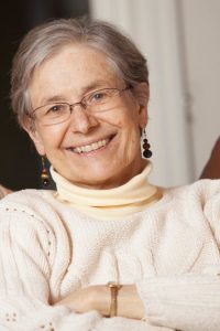
Our dear friend and colleague, Donata Oertel Sugden, PhD, passed away on April 22, 2020 from cancer. She was at home surrounded by her loving family.

Donata was a world renowned auditory neuroscientist whose discoveries of the role of cells in the cochlear nucleus will forever imprint our thinking about the neurophysiological basis of auditory function. She spent her career of 38 years at the University of Wisconsin-Madison, where she was until very recently Chair of the Neuroscience department. Donata was known to many members of ARO for the several decades that she was active in the association. Most notably, she was president of ARO in 2003, and Award of Merit recipient in 2004. She was a gentle, kind and brilliant woman. Her energy and enthusiasm were infectious and she was a role model for women scientists.

      • Please refer to the following link for an obituary published in Madison: LINK
      • And a tribute from the School of Medicine and Public Health at the University of Wisconsin Madison: LINK


For over 30 years Donata Oertel was the guiding light of neuroscience at the University of Wisconsin in Madison and contributed much to the university as a research scientist, a teacher and departmental chair. She received her undergraduate training at UC-Los Angeles and her Ph.D. in Biological Sciences from the UC-Santa Barbara. After post-doctoral research with Ching Kung in Madison, and with Ann Stuart at Harvard, she was appointed Assistant Professor of Neurophysiology at the University of Wisconsin-Madison in 1981 and was promoted to full Professor in 1992.

Donata Oertel began her independent research in 1981 as a new faculty member. After collaborating with Bill Rhode and Phil Smith to characterize the sound responses of neurons in the first relay station in the brain, the cochlear nuclear complex, in anesthetized cats, Dr. Oertel made the significant experimental leap of studying the nucleus using in vitro brain slices. At that time, not a lot was known about this nucleus because of its inaccessibility in the brain stem. In slices, neurons could be recorded and marked anatomically, and function could be explored with pharmacological tools. Use of the mammalian brain slice was a novel methodology in the 1980’s, but its application was restricted to a few brain regions such as the hippocampus, since other regions proved less amenable to long-term survival in vitro. So it was a major step to develop a brainstem slice preparation and excite neurons synaptically by stimulating the stump of the auditory nerve input. Introducing the cochlear nucleus brain slice enabled her to address a broad range of important questions about synaptic circuit mechanisms in sound processing. The premise was that to understand how auditory information is processed in the cochlear nuclei, it is crucial to know what circuitry exists and how it functions.

Her inaugural paper on the slice preparation (Oertel 1983) is a classic in the Journal of Neuroscience. It reveals a series of ultra-fast electrical responses that encode the key features of timing of auditory signals that must be key properties in sound localization. She was able to morphologically distinguish different cell types (bushy, stellate and octopus cells) with different firing patterns (Wu & Oertel 1984), and to record from the ventral cochlear nucleus (VCN) and the dorsal cochlear nucleus (DCN) (Hirsch & Oertel 1988; Zhang & Oertel 1993), regions of the early auditory pathway having distinct properties and physiological functions. Important findings include: (i) the delayed, frequency-specific inhibition from the DCN back to neurons in the VCN with properties suggesting it mediates suppression of echoes (Wickesberg & Oertel 1990); (ii) characterization of VCN octopus cells, that can detect with great precision coincident firing of auditory nerve inputs (Golding et al 1995; Ferragamo & Oertel 2002), and compensate for variable frequency-dependent delays in the cochlear traveling wave (McGinley et al 2012); (iii) the unusually fast kinetics of both voltage-dependent ion channels (Oertel 1997) and glutamate-receptor channels (Gardner et al 2001) that may be optimized for fast auditory signaling; (iv) demonstrating that multimodal sensory inputs on the dendrites of DCN fusiform cells were subject to long-term potentiation but that auditory inputs were not (Fujino & Oertel 2003). This multi-sensory convergence may compensate for head and pinna orientation during the sound localization process (Oertel & Young 2004). Work with her long-term collaborator Larry Trussell showed that long-term potentiation and depression, in the form of spike-timing dependent plasticity, were exhibited in the fusiform and cartwheel neurons of the DCN (Tzounopoulos et al. 2004); (v) the crucial roles of two membrane ion channels, the low-voltage activated K+ channel, Kv1.1 and hyperpolarization-activated cation channel, HCN1, which are co-regulated to set the neuronal resting potential. HCN1 in particular may be part of a process to detect gaps between acoustic stimuli (Cao & Oertel 2011; 2017; Oertel et al., 2020).

Donata Oertel always attempted to employ novel experimental approaches. One recent example was exploiting a mutation of receptor guanyl cyclase to understand the topographic organization of the auditory nerve input to the VCN (Lu et al 2014). Other examples include the involvement of nitric oxide signaling in potentiation between stellate cells tuned to the same frequency (Oertel & Cao 2017); and using an optogenetic technique, a hybrid voltage sensor, to explore synaptic connections between neurons excited with a pure tone (Lin et al 2018). Her work has touched on many features of neural signaling: optimizing response speed with fast glutamate receptors and voltage-dependent ion channels; long-term synaptic modification; differential sustained or transient firing patterns; dendritic integration and coincidence detection. Over the last 39 years, Donata Oertel has produced a body of work representing major advances in our understanding of how the brain processes and interprets sound. She was president of the Association for Research in Otolaryngology in 2002 and received the ARO Award of Merit in 2004. She served as a member of the NIDCD council (2003-2007) and was a member of the Board of Scientific Counselors, NIDCD (2011-2017). She delivered a plenary lecture at the Society for Neuroscience annual meeting in 2012.

Besides her research, Donata was a consummate teacher who contributed in no fewer than 18 disparate courses at UW Madison. She delivered lectures to a wide array of undergraduate, graduate and medical students covering an extraordinary range of subjects, ranging from brainstem auditory nuclei to the biophysical role of dendrites, from prions to brain cancers. She was kind and generous and always managed to pitch the lecture at a level appropriate for her audience. Donata also served in a number of administrative roles, and weathered challenging periods during departmental reorganizations and mergers. She served as chair of the Department of Neurophysiology, interim chair of the Department of Physiology and most recently as chair of the newly formed Department of Neuroscience, a position she occupied from 2014 until her death in spring 2020.

Donata is survived by her husband Bill Sugden, a virologist at UW-Madison, her son Arthur Sugden, and her daughter-in-law Lauren Sugden. Donata inspired Arthur to earn a Ph.D. in neuroscience, and they collaborated on several studies. Besides her loving family, Donata’s influence on the professional careers of countless colleagues stands as a legacy to her life and career.

Written by: Robert Fettiplace, Ph.D, University of Wisconsin-Madison

Hearing loss can significantly disrupt the ability of children to become mainstreamed in educational environments that emphasize spoken language as a primary means of communication. Similarly, adults who lose their hearing after communicating using spoken language have numerous challenges understanding speech and integrating into social situations. These challenges are particularly significant in noisy situations, where multiple sound sources often arrive at the ears from various directions. Intervention with hearing aids and/or cochlear implants (CIs) has proven to be highly successful for restoring some aspects of communication, including speech understanding and language acquisition. However, there is also typically a notable gap in outcomes relative to normal-hearing listeners. Importantly, auditory abilities operate in the context of how hearing integrates with other senses. Notably, the visual system is tightly couples to the auditory system. Vision is known to impact auditory perception and neural mechanisms in vision and audition are tightly coupled, thus, in order to understand how we hear and how CIs affect auditory perception we must consider the integrative effects across these senses.

We start with Rebecca Alexander, a compelling public speaker who has been living with Usher’s Syndrome, a genetic disorder found in tens of thousands of people, causing both deafness and blindness in humans. Ms. Alexander will be introduced by Dr. Jeffrey Holt, who studies gene therapy strategies for hearing restoration. The symposium then highlights the work of scientists working across these areas. Here we integrate psychophysics, clinical research, and biological approaches, aiming to gain a coherent understanding of how we might ultimately improve outcomes in patients. Drs. Susana Martinez-Conde and Stephen Macknik are new to the ARO community, and will discuss neurobiology of the visual system as it relates to visual prostheses. Dr. Jennifer Groh’s work will then discuss multi-sensory processing and how it is that vision helps us hear. Having set the stage for thinking about the role of vision in a multisensory auditory world, we will hear from experts in the area of cochlear implants. Dr. René H Gifford will discuss recent work on electric-acoustic integration in children and adults, and Dr. Sharon Cushing will discuss her work as a clinician on 3-D auditory and vestibular effects. Dr. Matthew Winn will talk about cognitive load and listening effort using pupillometry, and we will end with Dr. Rob Shepherd’s discussion of current work and future possibilities involving biological treatments and neural prostheses. Together, these presentations are designed to provide a broad and interdisciplinary view of the impact of sensory restoration in hearing, vision and balance, and the potential for future approaches for improving the lives of patients.

Kirupa Suthakar, PhD - Dr Kirupa Suthakar is a postdoctoral fellow at NIH/NIDCD, having formerly trained as a postdoctoral fellow at Massachusetts Eye and Ear/Harvard Medical School and doctoral student at Garvan Institute of Medical Research/UNSW Australia.  Kirupa's interest in the mind and particular fascination by how we are able to perceive the world around us led her to pursue a research career in auditory neuroscience.  To date, Kirupa's research has broadly focused on neurons within the auditory efferent circuit, which allow the brain to modulate incoming sound signals at the ear.  Kirupa is active member of the spARO community, serving as the Chair Elect for 2021.


I began studying the vestibular system during my dissertation research at the Università di Pavia with Professors Ivo Prigioni and GianCarlo Russo. I had two postdoctoral fellowships, first at the University of Rochester with Professor Christopher Holt and then at the University of Illinois at Chicago with Professors Jonathan Art and Jay Goldberg.

My research focuses on characterizing the biophysics of synaptic transmission between hair cells and primary afferents in the vestibular system. For many years an outstanding question in vestibular physiology was how the transduction current in the type I hair cell was sufficient, in the face of large conductances on at rest, to depolarize it to potentials necessary for conventional synaptic transmission with its unique afferent calyx.

In collaboration with Dr. Art, I overcame the technical challenges of simultaneously recording from type I hair cells and their enveloping calyx afferent to investigate this question. I was able to show that with depolarization of either hair cell or afferent, potassium ions accumulating in the cleft depolarize the synaptic partner. Conclusions from these studies are that due to the extended apposition between type I hair cell and its afferent, there are three modes of communication across the synapse. The slowest mode of transmission reflects the dynamic changes in potassium ion concentration in the cleft which follow the integral of the ongoing hair cell transduction current. The intermediate mode of transmission is indirectly a result of this potassium elevation which serves as the mechanism by which the hair cell potential is depolarized to levels necessary for calcium influx and the vesicle fusion typical of glutamatergic quanta. This increase in potassium concentration also depolarizes the afferent to potentials that allow the quantal EPSPs to trigger action potentials. The third and most rapid mode of transmission like the slow mode of transmission is bidirectional, and a current flowing out of either hair cell or afferent into the synaptic cleft will divide between a fraction flowing out into the bath, and a fraction flowing across the cleft into its synaptic partner.

The technical achievement of the dual electrode approach has enabled us to identify new facets of vestibular end organ synaptic physiology that in turn raise new questions and challenges for our field. I look forward with great excitement to the next chapter in my scientific story.


Charles C. Della Santina, PhD MD is a Professor of Otolaryngology – Head & Neck Surgery and Biomedical Engineering at the Johns Hopkins University School of Medicine, where he directs the Johns Hopkins Cochlear Implant Center and the Johns Hopkins Vestibular NeuroEngineering Laboratory.

As a practicing neurotologic surgeon, Dr. Della Santina specializes in treatment of middle ear, inner ear and auditory/vestibular nerve disorders. His clinical interests include restoration of hearing via cochlear implantation and management of patients who suffer from vestibular disorders, with a particular focus on helping individuals disabled by chronic postural instability and unsteady vision after bilateral loss of vestibular sensation. His laboratory’s research centers on basic and applied research supporting development of vestibular implants, which are medical devices intended to partially restore inner ear sensation of head movement. In addition to that work, his >90 publications include studies characterizing inner ear physiology and anatomy; describing novel clinical tests of vestibular function; and clarifying the effects of cochlear implantation, vestibular implantation, superior canal dehiscence syndrome and intratympanic gentamicin therapy on the inner ear and central nervous system.  Dr. Della Santina is also the founder and CEO/Chief Scientific Officer of Labyrinth Devices LLC, a company dedicated to bringing novel vestibular testing and implant technology into routine clinical care.

Andrew Griffith received his MD and PhD in Molecular Biophysics and Biochemistry from Yale University in 1992. He completed his general surgery internship and a residency in Otolaryngology-Head and Neck Surgery at the University of Michigan in 1998. He also completed a postdoctoral research fellowship in the Department of Human Genetics as part of his training at the University of Michigan. In 1998, he joined the Division of Intramural Research (DIR) in the National Institute on Deafness and Other Communication Disorders (NIDCD). He served as a senior investigator, the chief of the Molecular Biology and Genetics Section, the chief of the Otolaryngology Branch, and the director of the DIR, as well as the deputy director for Intramural Clinical Research across the NIH Intramural Research Program. His research program identifies and characterizes molecular and cellular mechanisms of normal and disordered hearing and balance in humans and mouse models. Two primary interests of his program have been hearing loss associated with enlargement of the vestibular aqueduct, and the function of TMC genes and proteins. The latter work lead to the discovery that the deafness gene product TMC1 is a component of the hair cell sensory transduction channel. Since July of 2020, he has served as the Senior Associate Dean of Research and a Professor of Otolaryngology and Physiology in the College of Medicine at the University of Tennessee Health Science Center.

Gwenaëlle S. G. Géléoc obtained a PhD in Sensory Neurobiology from the University of Sciences in Montpellier (France) in 1996. She performed part of her PhD training at the University of Sussex, UK where she characterized sensory transduction in vestibular hair cells and a performed a comparative study between vestibular and cochlear hair cells. Gwenaelle continued her training as an electrophysiologist at University College London studying outer hair cell motility and at Harvard Medical School studying modulation of mechanotransduction in vestibular hair cells. As an independent investigator at the University of Virginia, she expanded this work and characterized the developmental acquisition of sensory transduction in mouse vestibular hair cells, the developmental acquisition of voltage-sensitive conductances in vestibular hair cells and the tonotopic gradient in the acquisition of sensory transduction in the mouse cochlea. This work along with quantitative spatio-temporal studies performed on several hair cell mechanotransduction candidates lead her to TMC1 and 2 and long-term collaborations with Andrew Griffith and Jeff Holt. Dr. Géléoc is currently Assistant Professor of Otolaryngology, at Boston Children’s Hospital where she continues to study molecular players involved in the development and function of hair cells of the inner ear and develops new therapies for the treatment of deafness and balance, with a particular focus on Usher syndrome.

Jeff Holt earned a doctorate from the Department of Physiology at the University of Rochester in 1995 for his studies of inward rectifier potassium channels in saccular hair cells.  He went on to a post-doctoral position in the Neurobiology Department at Harvard Medical School and the Howard Hughes Medical Institute, where he characterized sensory transduction and adaptation in hair cells and developed a viral vector system to transfect cultured hair cells.  Dr. Holt’s first faculty position was in the Neuroscience Department at the University of Virginia.  In 2011 the lab moved to Boston Children’s Hospital / Harvard Medical School.  Dr. Holt is currently a Professor in the Departments of Otolaryngology and Neurology in the F.M. Kirby Neurobiology Center.  Dr. Holt and his team have been studying sensory transduction in auditory and vestibular hair cells over the past 20 years, with particular focus on TMC1 and TMC2 over the past 12 years.  This work lead to the discovery that TMC1 forms the hair cell transduction channel.  His work also focuses on development gene therapy strategies for genetic hearing loss.